Glaucoma Clinic
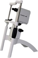
- Tonometry
- Automated Perimetry
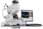
- HRT-II
- Gonioscopy
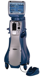
- Glaucoma Surgery
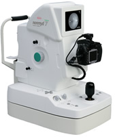
At our centre we have the facilities of diagnosing and treating the patients of glaucoma.  Glaucoma is a disease in which the pressure inside the eye rises. This raised pressure tends to damage the nerve of the eye (optic nerve). We have the facilities of Intraocular pressure recording by computerized and non-computerized methods both.